What is a Carotid Doppler Ultrasound?
A carotid Doppler ultrasound is a safe, noninvasive test. Doctors use it to check the blood flow in your neck’s carotid arteries. These arteries carry blood from your heart to your brain. Because blockages here can lead to strokes, this test is important for stroke prevention imaging. Many hospitals and clinics in your area offer this test.
How the Test Works
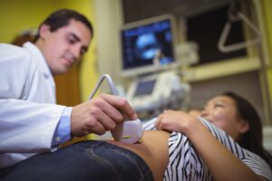
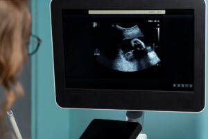
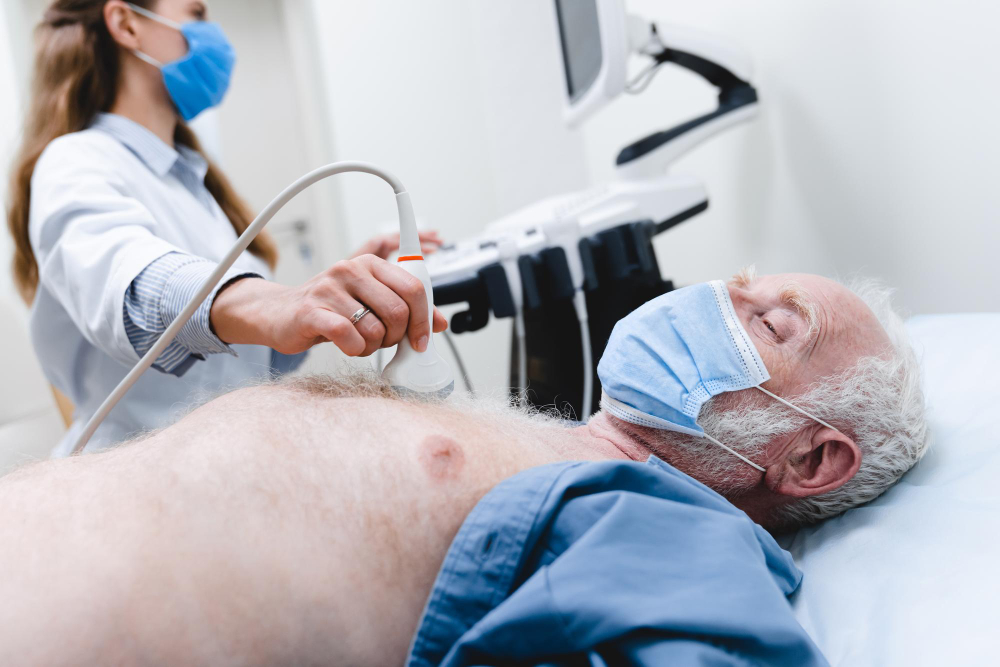
During the test, a small device called a transducer moves over your neck. It sends sound waves into your body. Then, these waves bounce off your blood vessels. The machine turns these echoes into pictures. As a result, doctors can see if your arteries are blocked or narrowed. This noninvasive carotid artery test does not use needles or radiation.
Why It’s Used to Assess Stroke Risk
Doctors use a carotid Doppler ultrasound for stroke risk assessment. If your carotid arteries are blocked, blood may not reach your brain. This can cause a stroke. However, finding blockages early can help prevent strokes. According to the CDC, stroke is a leading cause of death and disability. That is why early detection is so important.
Symptoms and Risk Factors That May Lead to Testing
Sometimes, people do not notice any symptoms. Still, doctors may suggest this test if you have certain risks. For example, you may need a carotid Doppler ultrasound if you have:
Additionally, symptoms such as sudden weakness, trouble speaking, or vision changes may lead to testing. Early testing can help lower your risk of stroke.
How to Prepare for the Procedure
For most people, no special steps are needed before a carotid Doppler ultrasound. However, your doctor may ask you to avoid wearing necklaces or clothing with tight collars. This makes it easier to reach your neck. You can eat and take your usual medicines unless your doctor says otherwise. If you have questions, ask your healthcare team before the test.
What to Expect During and After the Test
First, you will lie down on an exam table. Next, a technician will put a small amount of gel on your neck. This gel helps the sound waves travel better. Then, the technician moves the transducer over your skin. You may hear a whooshing sound, which is your blood moving. The test usually takes 30 minutes or less. Afterward, the gel is wiped off, and you can return to normal activities right away.
Understanding the Results
After the test, a doctor reviews the images. If your arteries are clear, your stroke risk is lower. However, if there is narrowing or blockage, your doctor will talk with you about next steps. Sometimes, more tests or treatments are needed. Early treatment can help prevent a stroke. Always ask your doctor to explain your results in simple terms.
Prevention and Lifestyle Guidance Related to Stroke Risk
Even if your carotid Doppler ultrasound shows no blockage, it is wise to lower your stroke risk. For example, you can:
Because stroke prevention imaging helps find problems early, following your doctor’s advice is key. The World Health Organization and CDC both support healthy lifestyle changes to reduce stroke risk.